DSSR-enabled G4 identification/annotation: PDB entry 352d
Summary information and primary citation
- PDB-id
- 352d
- Class
- DNA
- Method
- X-ray (0.95 Å)
- Summary
- The crystal structure of a parallel-stranded parallel-stranded guanine tetraplex at 0.95 angstrom resolution
- Reference
- Phillips K, Dauter Z, Murchie AI, Lilley DM, Luisi B (1997): "The crystal structure of a parallel-stranded guanine tetraplex at 0.95 A resolution." J.Mol.Biol., 273, 171-182. doi: 10.1006/jmbi.1997.1292.
- Abstract
- In both DNA and RNA, stretches of guanine bases can form stable four-stranded helices in the presence of sodium or potassium ions. Sequences with a propensity to form guanine tetraplexes have been found in chromosomal telomers, immunoglobulin switch regions, and recombination sites. We report the crystal structure at 0.95 A resolution of a parallel-stranded tetraplex formed by the hexanucleotide d(TG4T) in the presence of sodium ions. The four strands form a right-handed helix that is stabilized by hydrogen-bonding tetrads of co-planar guanine bases. Well-resolved sodium ions are found between and, at defined points, within tetrad planes and are coordinated with the guanine O6 groups. Nine calcium ions have been identified, each with a well-defined hepta-coordinate hydration shell. Hydrogen-bonding water patterns are observed within the tetraplex's helical grooves and clustered about the phosphate groups. Water molecules in the groove may form a hydrogen bond with the O4', and may affect the stacking behavior of guanine. Two distinct stacking arrangements are noted for the guanine tetrads. The thymine bases do not contribute to the four-stranded conformation, but instead stack to stabilize the crystal lattice. We present evidence that the sugar conformation is strained and propose that this originates from forces that optimize guanine base stacking. Discrete conformational disorder is observed at several places in the phosphodiester backbone, which results from a simple crankshaft rotation that requires no net change in the sugar conformation.
- G4 notes
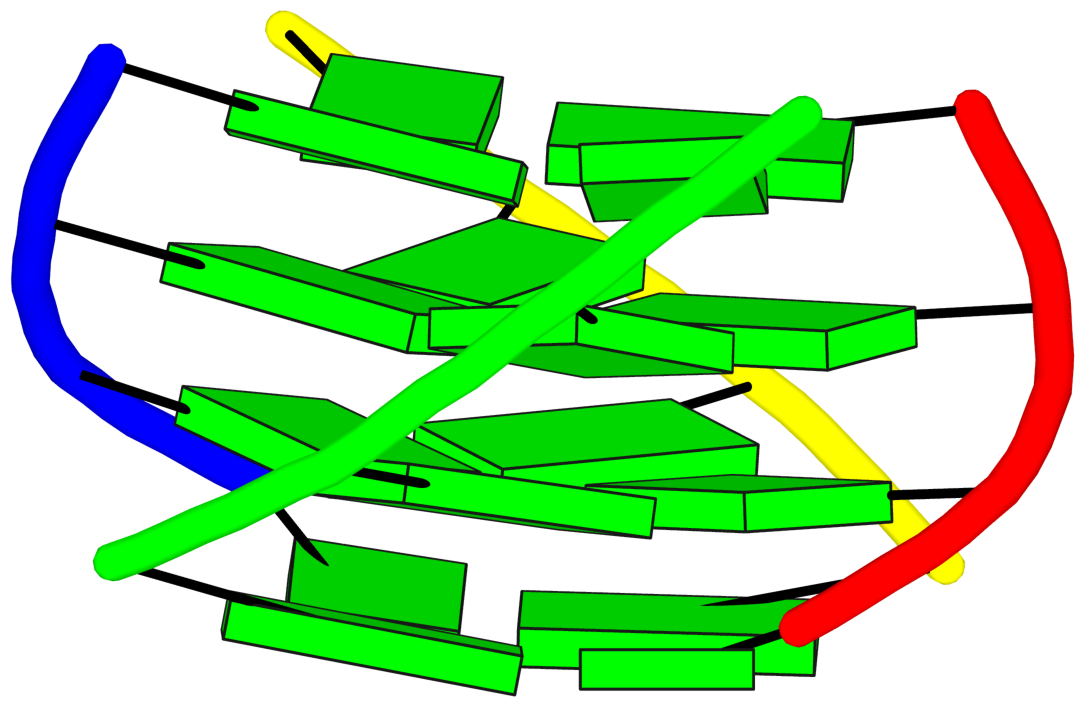
- 4 G-tetrads, 1 G4 helix, 1 G4 stem, UUUU, parallel, 4+0
Cartoon-block schematics in six views (download the tarball)
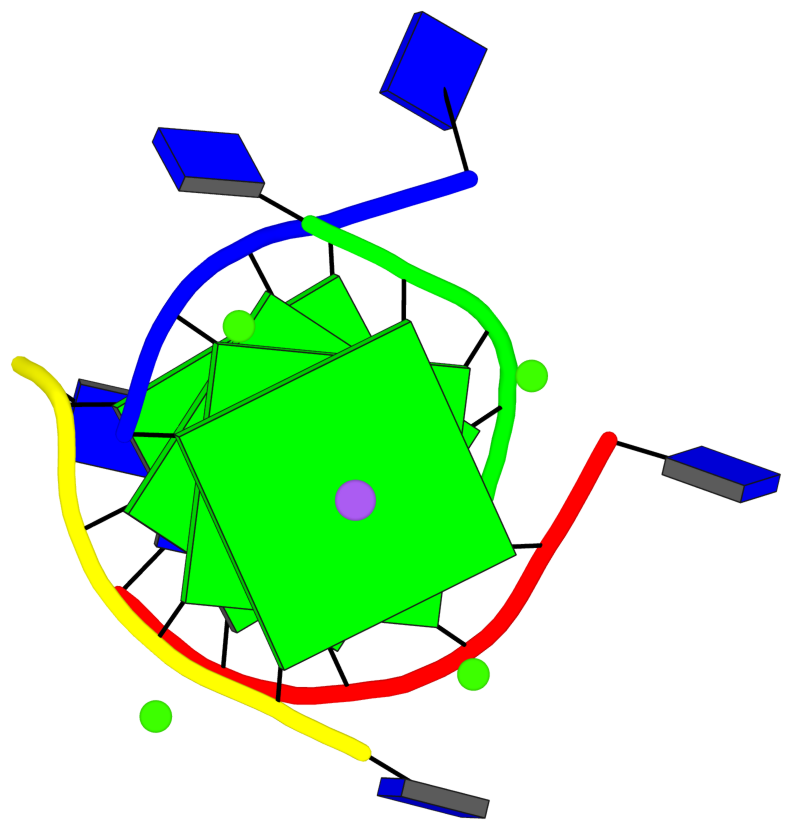
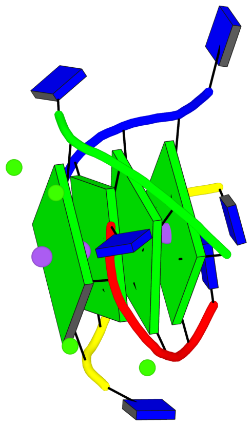
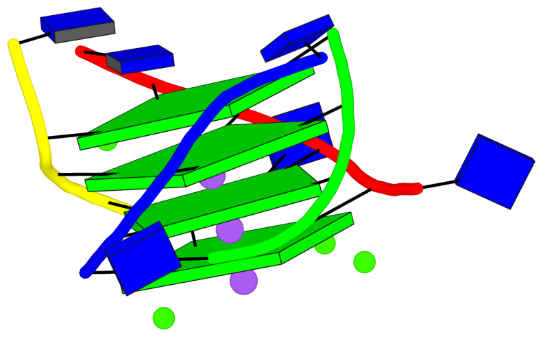
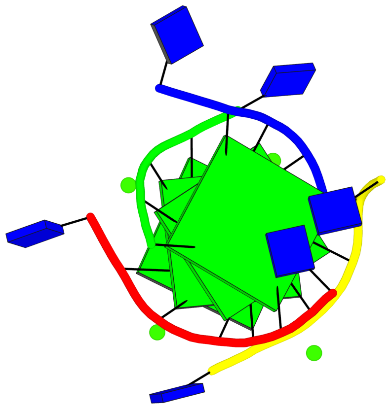
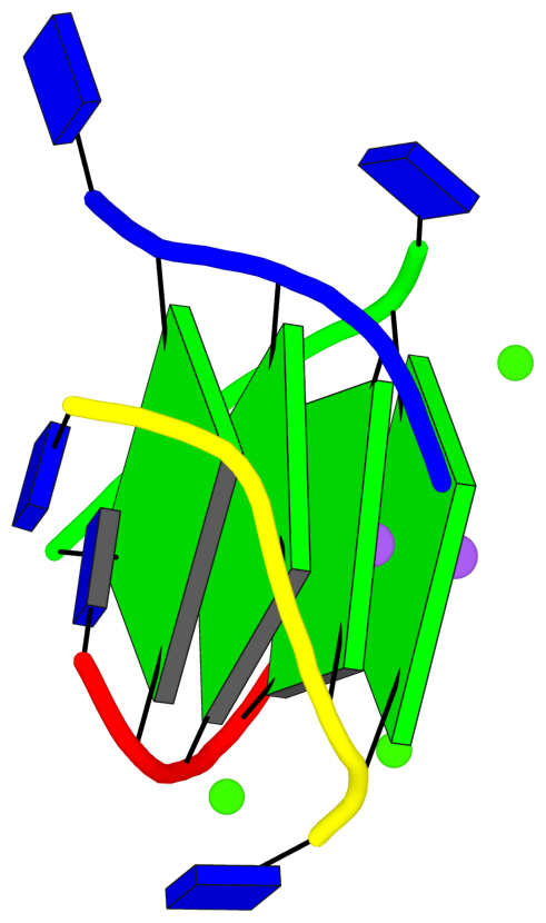
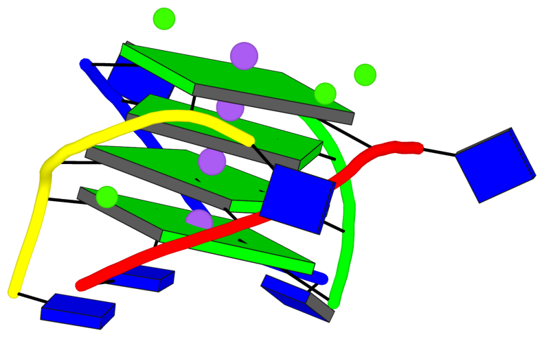
List of 4 G-tetrads
 1 glyco-bond=---- sugar=---- groove=---- planarity=0.224 type=bowl   -- nts=4 GGGG 1:A.DG2,1:D.DG32,1:C.DG22,1:B.DG12
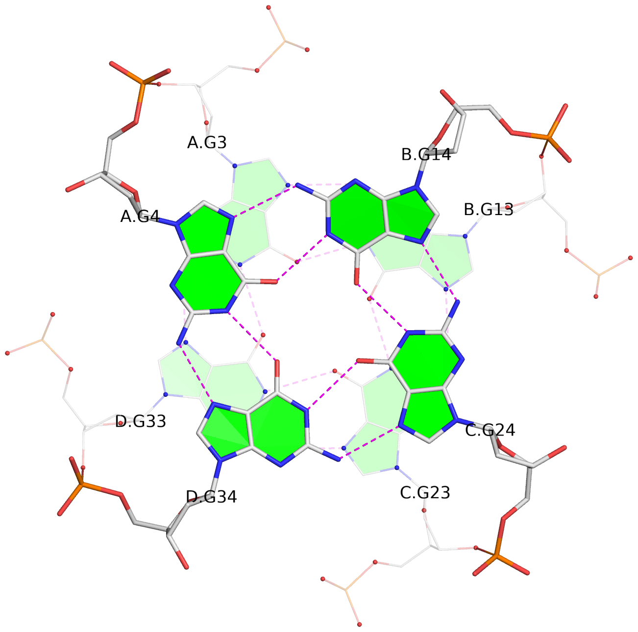
 2 glyco-bond=---- sugar=---- groove=---- planarity=0.210 type=bowl   -- nts=4 GGGG 1:A.DG3,1:D.DG33,1:C.DG23,1:B.DG13
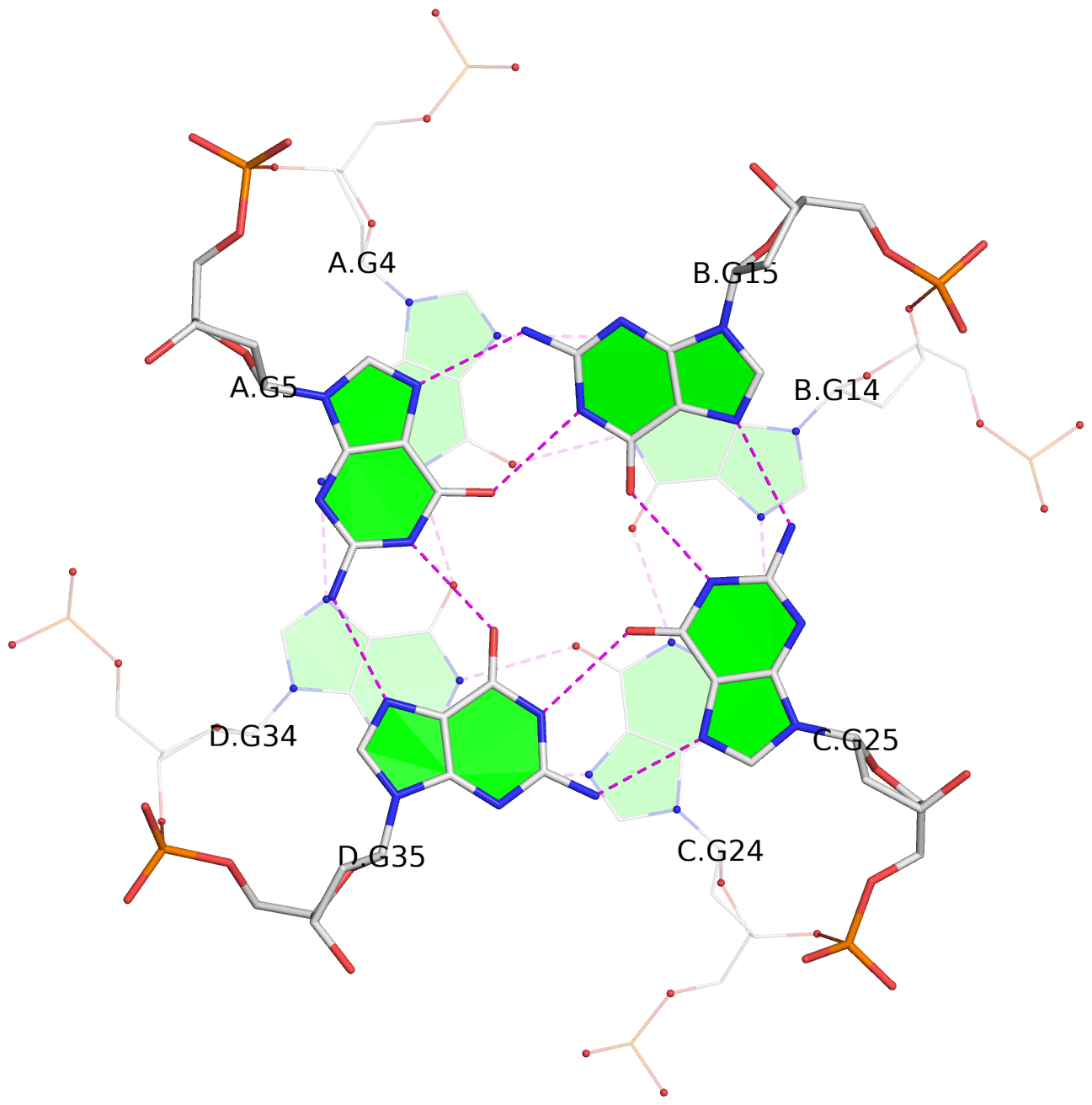
 3 glyco-bond=---- sugar=---- groove=---- planarity=0.257 type=bowl   -- nts=4 GGGG 1:A.DG4,1:D.DG34,1:C.DG24,1:B.DG14
 4 glyco-bond=---- sugar=---- groove=---- planarity=0.306 type=bowl   -- nts=4 GGGG 1:A.DG5,1:D.DG35,1:C.DG25,1:B.DG15
List of 1 G4-helix
In DSSR, a G4-helix is defined by stacking interactions of G-tetrads, regardless of backbone connectivity, and may contain more than one G4-stem.
Helix#1, 4 G-tetrads, inter-molecular, with 1 stem
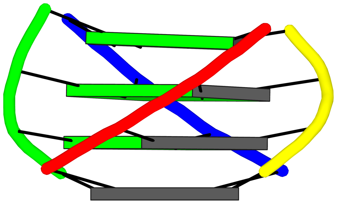
|
1 glyco-bond=---- sugar=---- groove=---- WC-->Major -- nts=4 GGGG 1:A.DG2,1:D.DG32,1:C.DG22,1:B.DG12 2 glyco-bond=---- sugar=---- groove=---- WC-->Major -- nts=4 GGGG 1:A.DG3,1:D.DG33,1:C.DG23,1:B.DG13 3 glyco-bond=---- sugar=---- groove=---- WC-->Major -- nts=4 GGGG 1:A.DG4,1:D.DG34,1:C.DG24,1:B.DG14 4 glyco-bond=---- sugar=---- groove=---- WC-->Major -- nts=4 GGGG 1:A.DG5,1:D.DG35,1:C.DG25,1:B.DG15 step#1 pm(>>,forward) area=8.82 rise=3.31 twist=31.7 step#2 pm(>>,forward) area=12.34 rise=3.29 twist=26.7 step#3 pm(>>,forward) area=11.62 rise=3.37 twist=27.8 strand#1 DNA glyco-bond=---- sugar=---- nts=4 GGGG 1:A.DG2,1:A.DG3,1:A.DG4,1:A.DG5 strand#2 DNA glyco-bond=---- sugar=---- nts=4 GGGG 1:D.DG32,1:D.DG33,1:D.DG34,1:D.DG35 strand#3 DNA glyco-bond=---- sugar=---- nts=4 GGGG 1:C.DG22,1:C.DG23,1:C.DG24,1:C.DG25 strand#4 DNA glyco-bond=---- sugar=---- nts=4 GGGG 1:B.DG12,1:B.DG13,1:B.DG14,1:B.DG15 |
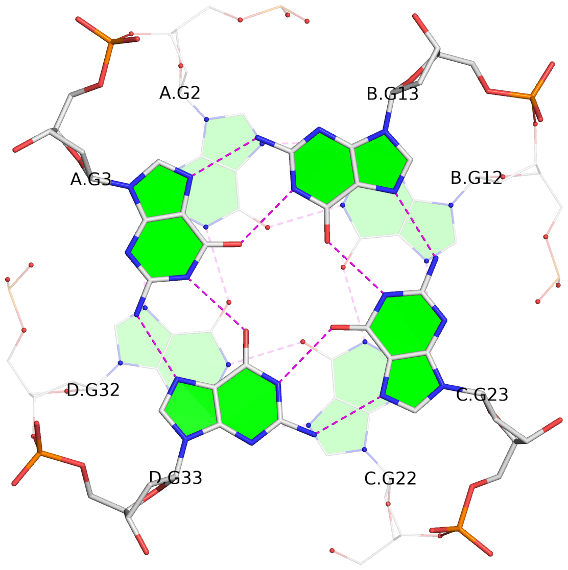
| 3 stacking diagrams | |
|
1 glyco-bond=---- sugar=---- groove=---- WC-->Major -- nts=4 GGGG 1:A.DG2,1:D.DG32,1:C.DG22,1:B.DG12 |
|
2 glyco-bond=---- sugar=---- groove=---- WC-->Major -- nts=4 GGGG 1:A.DG3,1:D.DG33,1:C.DG23,1:B.DG13 |
|
3 glyco-bond=---- sugar=---- groove=---- WC-->Major -- nts=4 GGGG 1:A.DG4,1:D.DG34,1:C.DG24,1:B.DG14 |
List of 1 G4-stem
In DSSR, a G4-stem is defined as a G4-helix with backbone connectivity. Bulges are also allowed along each of the four strands.
Stem#1, 4 G-tetrads, 0 loops, inter-molecular, UUUU, parallel, 4+0
|
1 glyco-bond=---- sugar=---- groove=---- WC-->Major -- nts=4 GGGG 1:A.DG2,1:D.DG32,1:C.DG22,1:B.DG12 2 glyco-bond=---- sugar=---- groove=---- WC-->Major -- nts=4 GGGG 1:A.DG3,1:D.DG33,1:C.DG23,1:B.DG13 3 glyco-bond=---- sugar=---- groove=---- WC-->Major -- nts=4 GGGG 1:A.DG4,1:D.DG34,1:C.DG24,1:B.DG14 4 glyco-bond=---- sugar=---- groove=---- WC-->Major -- nts=4 GGGG 1:A.DG5,1:D.DG35,1:C.DG25,1:B.DG15 step#1 pm(>>,forward) area=8.82 rise=3.31 twist=31.7 step#2 pm(>>,forward) area=12.34 rise=3.29 twist=26.7 step#3 pm(>>,forward) area=11.62 rise=3.37 twist=27.8 strand#1 U DNA glyco-bond=---- sugar=---- nts=4 GGGG 1:A.DG2,1:A.DG3,1:A.DG4,1:A.DG5 strand#2 U DNA glyco-bond=---- sugar=---- nts=4 GGGG 1:D.DG32,1:D.DG33,1:D.DG34,1:D.DG35 strand#3 U DNA glyco-bond=---- sugar=---- nts=4 GGGG 1:C.DG22,1:C.DG23,1:C.DG24,1:C.DG25 strand#4 U DNA glyco-bond=---- sugar=---- nts=4 GGGG 1:B.DG12,1:B.DG13,1:B.DG14,1:B.DG15 |