DSSR-enabled G4 identification/annotation: PDB entry 3qxr
Summary information and primary citation
- PDB-id
- 3qxr
- Class
- DNA
- Method
- X-ray (1.62 Å)
- Summary
- Crystal structure of the brominated ckit-1 proto-oncogene promoter quadruplex DNA
- Reference
- Wei D, Parkinson GN, Reszka AP, Neidle S (2012): "Crystal structure of a c-kit promoter quadruplex reveals the structural role of metal ions and water molecules in maintaining loop conformation." Nucleic Acids Res., 40, 4691-4700. doi: 10.1093/nar/gks023.
- Abstract
- We report here the 1.62 Å crystal structure of an intramolecular quadruplex DNA formed from a sequence in the promoter region of the c-kit gene. This is the first reported crystal structure of a promoter quadruplex and the first observation of localized magnesium ions in a quadruplex structure. The structure reveals that potassium and magnesium ions have an unexpected yet significant structural role in stabilizing particular quadruplex loops and grooves that is distinct from but in addition to the role of potassium ions in the ion channel at the centre of all quadruplex structures. The analysis also shows how ions cluster together with structured water molecules to stabilize the quadruplex arrangement. This particular quadruplex has been previously studied by NMR methods, and the present X-ray structure is in accord with the earlier topology assignment. However, as well as the observations of potassium and magnesium ions, the crystal structure has revealed a highly significant difference in the dimensions of the large cleft in the structure, which is a plausible target for small molecules. This difference can be understood by the stabilizing role of structured water networks.
- G4 notes
- 3 G-tetrads, 1 G4 helix, 1 G4 stem, UUUU, parallel, 4+0, 2(-PX+P)
Cartoon-block schematics in six views (download the tarball)
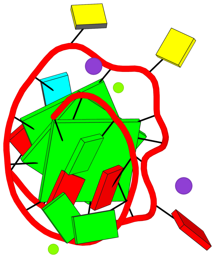
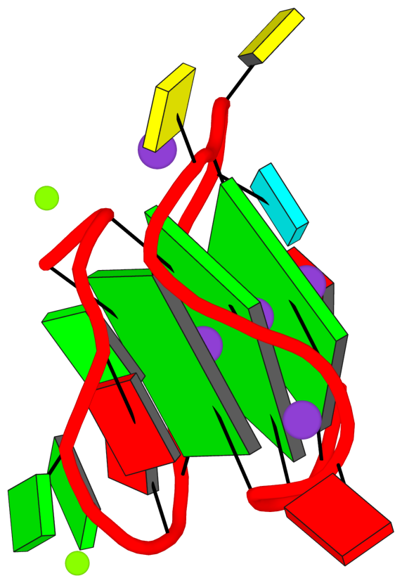
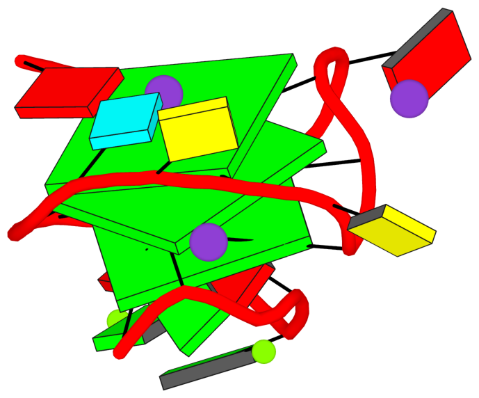
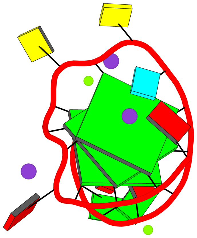
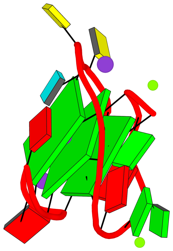
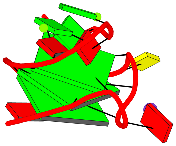
List of 3 G-tetrads
 1 glyco-bond=---- sugar=-3-- groove=---- planarity=0.230 type=other  O+ nts=4 GGGG 1:A.DG2,1:A.DG6,1:A.DG10,1:A.DG13
 2 glyco-bond=---- sugar=.-3- groove=---- planarity=0.272 type=other  N+ nts=4 GGGG 1:A.DG3,1:A.DG7,1:A.DG21,1:A.DG14
 3 glyco-bond=---- sugar=---- groove=---- planarity=0.393 type=bowl   N+ nts=4 GGGG 1:A.DG4,1:A.DG8,1:A.DG22,1:A.DG15
List of 1 G4-helix
In DSSR, a G4-helix is defined by stacking interactions of G-tetrads, regardless of backbone connectivity, and may contain more than one G4-stem.
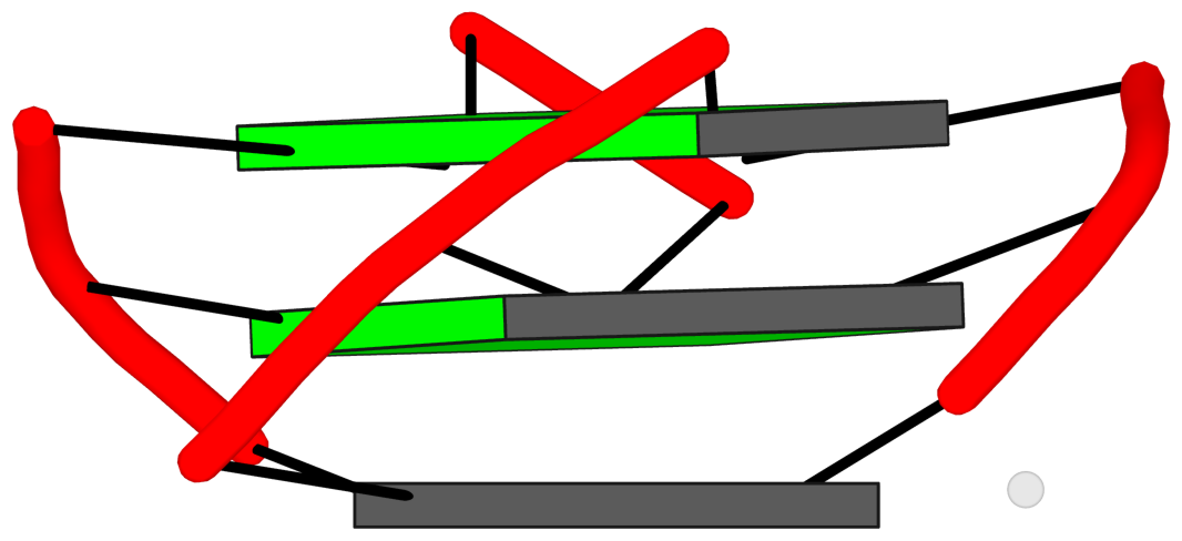
Helix#1, 3 G-tetrads, INTRA-molecular, with 1 stem
|
1 glyco-bond=---- sugar=-3-- groove=---- WC-->Major O+ nts=4 GGGG 1:A.DG2,1:A.DG6,1:A.DG10,1:A.DG13 2 glyco-bond=---- sugar=.-3- groove=---- WC-->Major N+ nts=4 GGGG 1:A.DG3,1:A.DG7,1:A.DG21,1:A.DG14 3 glyco-bond=---- sugar=---- groove=---- WC-->Major N+ nts=4 GGGG 1:A.DG4,1:A.DG8,1:A.DG22,1:A.DG15 step#1 pm(>>,forward) area=10.90 rise=3.40 twist=29.0 step#2 pm(>>,forward) area=8.55 rise=3.39 twist=32.5 strand#1 DNA glyco-bond=--- sugar=-.- nts=3 GGG 1:A.DG2,1:A.DG3,1:A.DG4 strand#2 DNA glyco-bond=--- sugar=3-- nts=3 GGG 1:A.DG6,1:A.DG7,1:A.DG8 strand#3 DNA glyco-bond=--- sugar=-3- nts=3 GGG 1:A.DG10,1:A.DG21,1:A.DG22 strand#4 DNA glyco-bond=--- sugar=--- nts=3 GGG 1:A.DG13,1:A.DG14,1:A.DG15 |
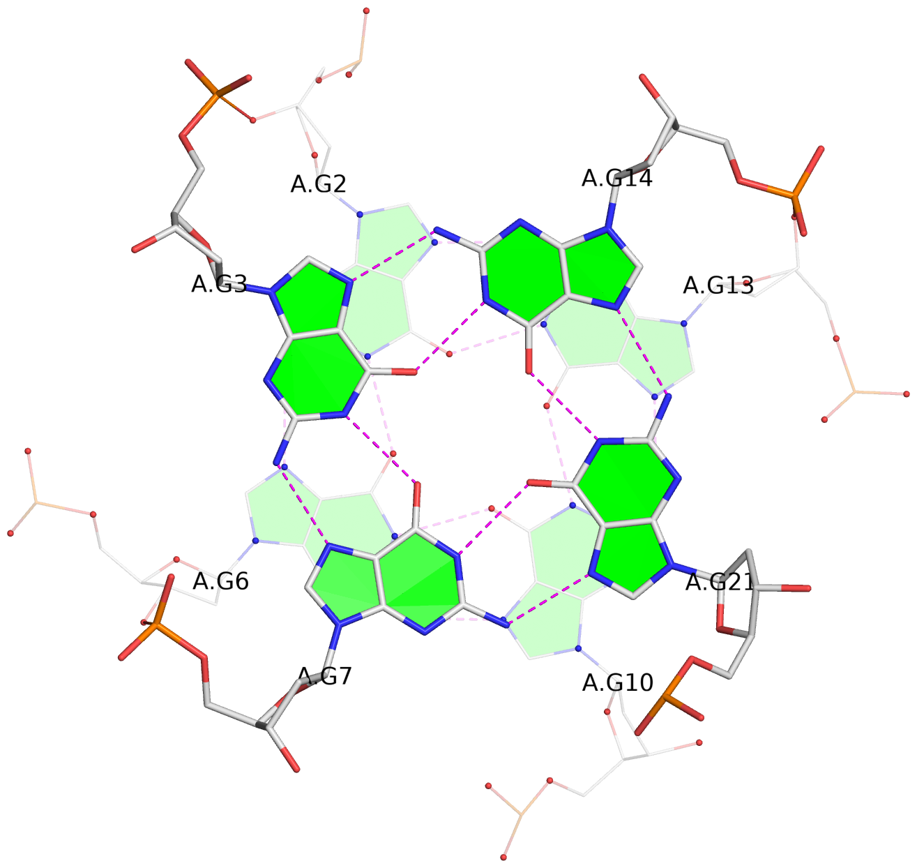
| 2 stacking diagrams | |
|
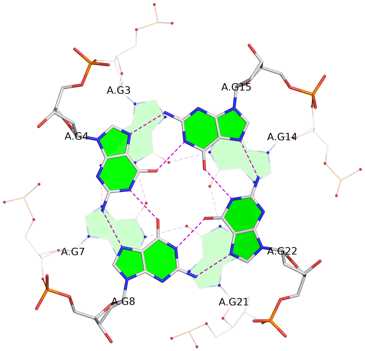
1 glyco-bond=---- sugar=-3-- groove=---- WC-->Major O+ nts=4 GGGG 1:A.DG2,1:A.DG6,1:A.DG10,1:A.DG13 |
|
2 glyco-bond=---- sugar=.-3- groove=---- WC-->Major N+ nts=4 GGGG 1:A.DG3,1:A.DG7,1:A.DG21,1:A.DG14 |
List of 1 G4-stem
In DSSR, a G4-stem is defined as a G4-helix with backbone connectivity. Bulges are also allowed along each of the four strands.
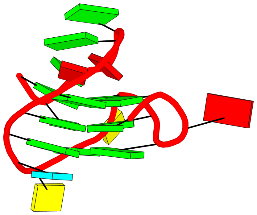
Stem#1, 2 G-tetrads, 3 loops, INTRA-molecular, UUUU, parallel, 4+0, 2(-PX+P)
|
1 glyco-bond=---- sugar=.-3- groove=---- WC-->Major N+ nts=4 GGGG 1:A.DG3,1:A.DG7,1:A.DG21,1:A.DG14 2 glyco-bond=---- sugar=---- groove=---- WC-->Major N+ nts=4 GGGG 1:A.DG4,1:A.DG8,1:A.DG22,1:A.DG15 step#1 pm(>>,forward) area=8.55 rise=3.39 twist=32.5 strand#1 U DNA glyco-bond=-- sugar=.- nts=2 GG 1:A.DG3,1:A.DG4 strand#2 U DNA glyco-bond=-- sugar=-- nts=2 GG 1:A.DG7,1:A.DG8 strand#3 U DNA glyco-bond=-- sugar=3- nts=2 GG 1:A.DG21,1:A.DG22 strand#4 U DNA glyco-bond=-- sugar=-- nts=2 GG 1:A.DG14,1:A.DG15 loop#1 type=propeller strands=[#1,#2] nts=2 AG 1:A.DA5,1:A.DG6 loop#2 type=diag-prop strands=[#2,#4] nts=5 CGCuG 1:A.DC9,1:A.DG10,1:A.DC11,1:A.BRU12,1:A.DG13 loop#3 type=propeller strands=[#4,#3] nts=5 AGGAG 1:A.DA16,1:A.DG17,1:A.DG18,1:A.DA19,1:A.DG20 |
List of 2 non-stem G4-loops (including the two closing Gs)
 1* type=V-shaped  helix=#1 nts=5 GGGCG 1:A.DG6,1:A.DG7,1:A.DG8,1:A.DC9,1:A.DG10
 2 type=lateral   helix=#1 nts=4 GCuG 1:A.DG10,1:A.DC11,1:A.BRU12,1:A.DG13