DSSR-enabled G4 identification/annotation: PDB entry 5yey
Summary information and primary citation
- PDB-id
- 5yey
- Class
- DNA
- Method
- NMR
- Summary
- The structure of a chair-type g-quadruplex of the human telomeric variant in k+ solution
- Reference
- Liu C, Zhou B, Geng Y, Yan Tam D, Feng R, Miao H, Xu N, Shi X, You Y, Hong Y, Tang BZ, Kwan Lo P, Kuryavyi V, Zhu G (2019): "A chair-type G-quadruplex structure formed by a human telomeric variant DNA in K+solution." Chem Sci, 10, 218-226. doi: 10.1039/c8sc03813a.
- Abstract
- Guanine tracts of human telomeric DNA sequences are known to fold into eight different four-stranded structures that vary by the conformation of guanine nucleotides arranged in the stack of G-tetrads in their core and by different kinds and orders of connecting loops, called G-quadruplexes. Here, we present a novel G-quadruplex structure formed in K<sub>+</sub> solution by a human telomeric variant d[(GGGTTA)2GGGTTTGGG], <i>htel21</i>T<sub>18</sub>. This variant DNA is located in the subtelomeric regions of human chromosomes 8, 11, 17, and 19 as well as in the DNase hypersensitive region and in the subcentromeric region of chromosome 5. Interestingly, single A18T substitution that makes <i>htel21</i>T<sub>18</sub> different from the human telomeric sequence results in the formation of a three-layer chair-type G-quadruplex, a fold previously unknown among human telomeric repeats, with two loops interacting through the reverse Watson-Crick A6·T18 base pair. The loops are edgewise; glycosidic conformation of guanines is <i>syn</i>·<i>anti</i>·<i>syn</i>·<i>anti</i> around each tetrad, and each strand of the core has two antiparallel adjacent strands. Our results expand the repertoire of known G-quadruplex folding topologies and may provide a potential target for structure-based anticancer drug design.
- G4 notes
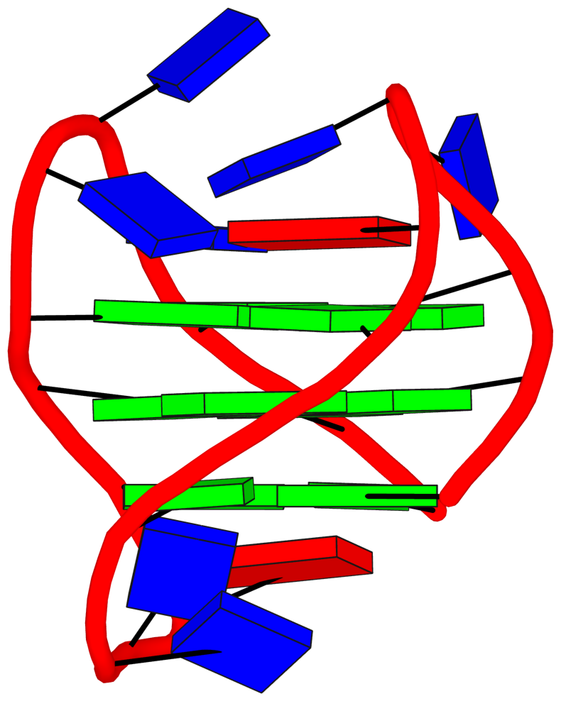
- 3 G-tetrads, 1 G4 helix, 1 G4 stem, UDUD, anti-parallel:chair, 2+2, 3(+Ln+Lw+Ln)
Cartoon-block schematics in six views (download the tarball)
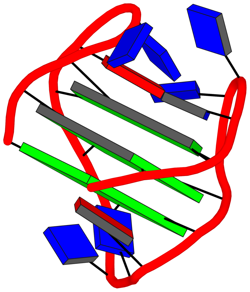
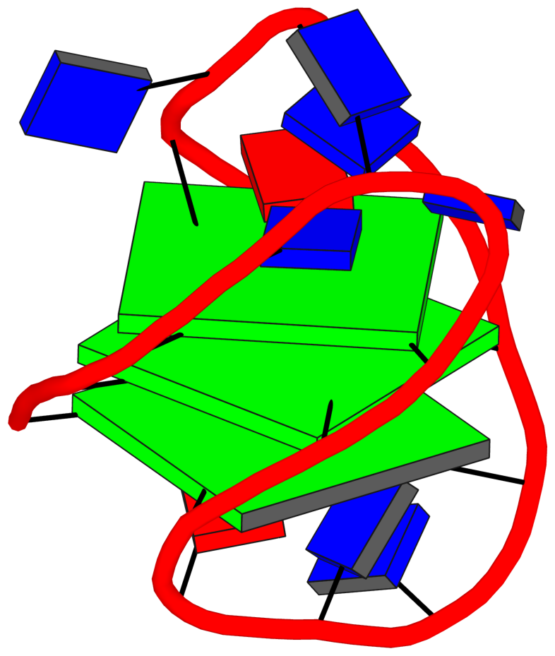
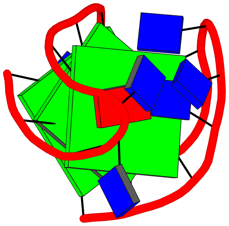
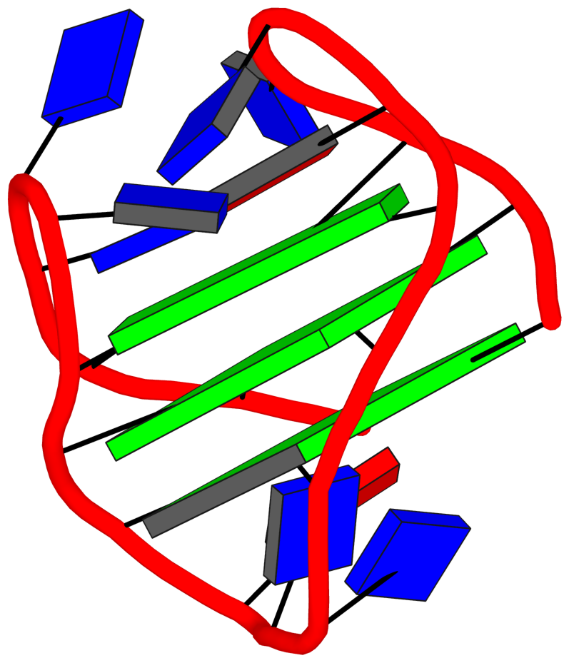
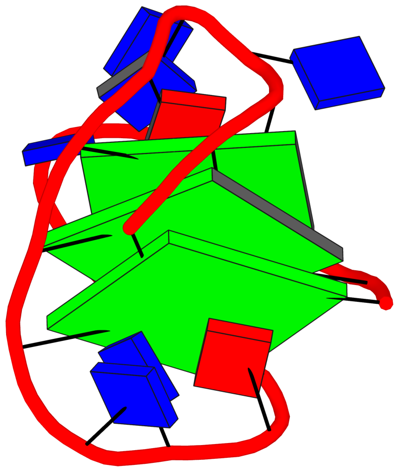
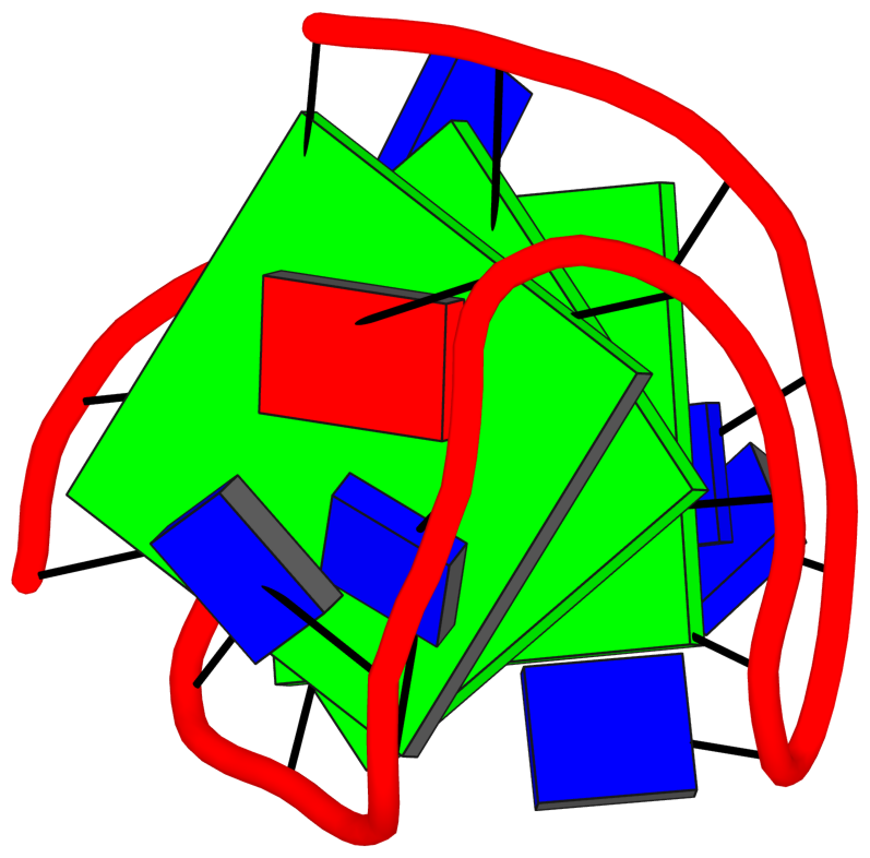
List of 3 G-tetrads
 1 glyco-bond=s-s- sugar=-.-- groove=wnwn planarity=0.080 type=planar O+ nts=4 GGGG A.DG1,A.DG21,A.DG13,A.DG9
 2 glyco-bond=-s-s sugar=---3 groove=wnwn planarity=0.010 type=planar O- nts=4 GGGG A.DG2,A.DG20,A.DG14,A.DG8
 3 glyco-bond=-s-s sugar=3--- groove=wnwn planarity=0.063 type=planar O- nts=4 GGGG A.DG3,A.DG19,A.DG15,A.DG7
List of 1 G4-helix
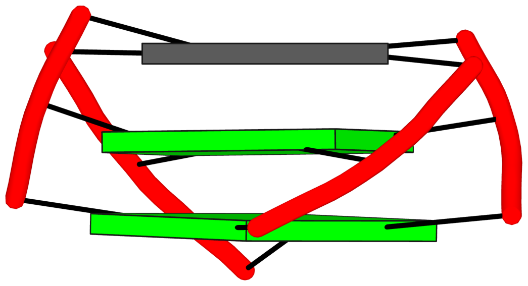
In DSSR, a G4-helix is defined by stacking interactions of G-tetrads, regardless of backbone connectivity, and may contain more than one G4-stem.
Helix#1, 3 G-tetrads, INTRA-molecular, with 1 stem
|
1 glyco-bond=s-s- sugar=-.-- groove=wnwn Major-->WC O+ nts=4 GGGG A.DG1,A.DG21,A.DG13,A.DG9 2 glyco-bond=-s-s sugar=---3 groove=wnwn WC-->Major O- nts=4 GGGG A.DG2,A.DG20,A.DG14,A.DG8 3 glyco-bond=-s-s sugar=3--- groove=wnwn WC-->Major O- nts=4 GGGG A.DG3,A.DG19,A.DG15,A.DG7 step#1 mm(<>,outward) area=12.07 rise=3.46 twist=18.4 step#2 pm(>>,forward) area=9.90 rise=3.34 twist=32.3 strand#1 DNA glyco-bond=s-- sugar=--3 nts=3 GGG A.DG1,A.DG2,A.DG3 strand#2 DNA glyco-bond=-ss sugar=.-- nts=3 GGG A.DG21,A.DG20,A.DG19 strand#3 DNA glyco-bond=s-- sugar=--- nts=3 GGG A.DG13,A.DG14,A.DG15 strand#4 DNA glyco-bond=-ss sugar=-3- nts=3 GGG A.DG9,A.DG8,A.DG7 |
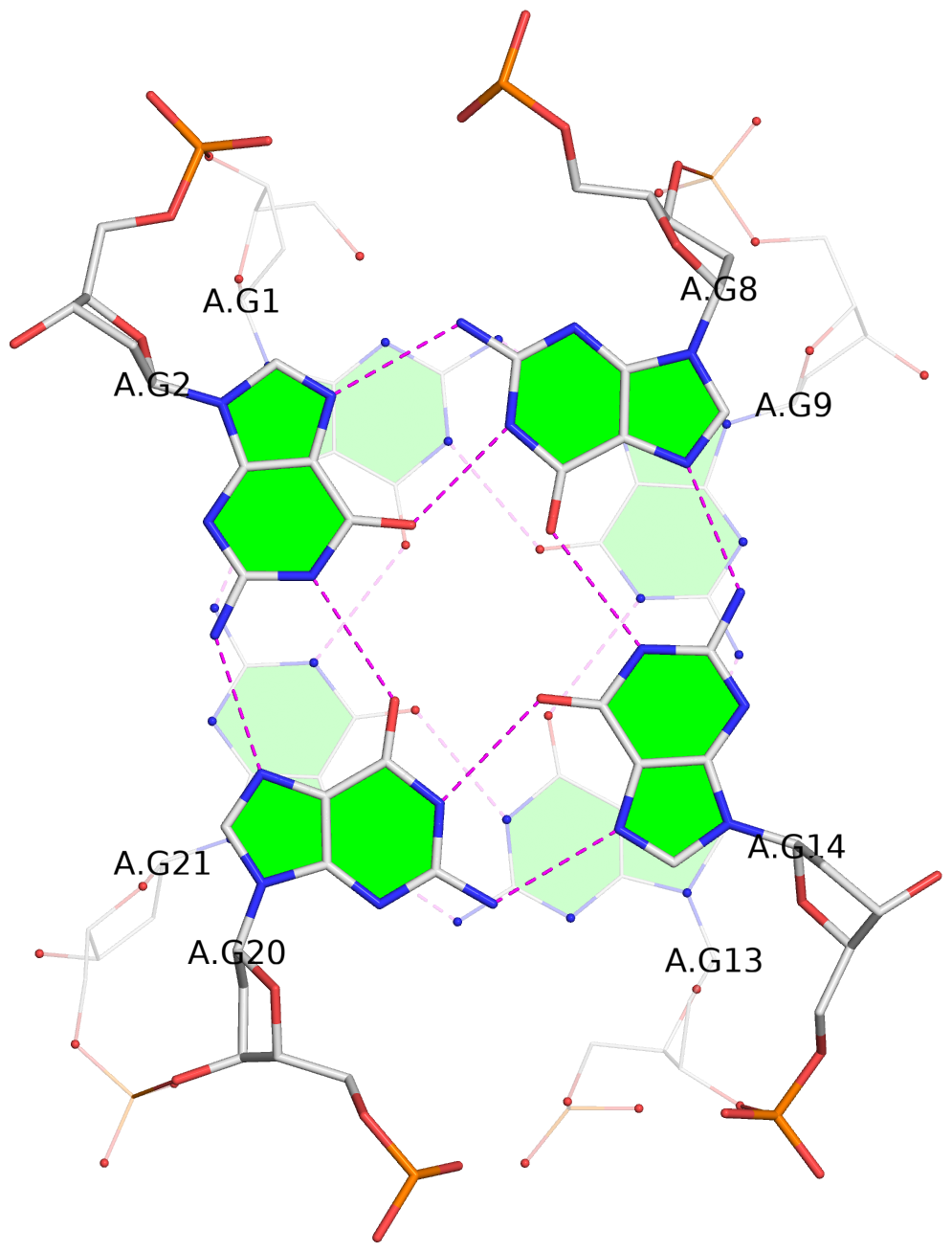
| 2 stacking diagrams | |
|
1 glyco-bond=s-s- sugar=-.-- groove=wnwn Major-->WC O+ nts=4 GGGG A.DG1,A.DG21,A.DG13,A.DG9 |
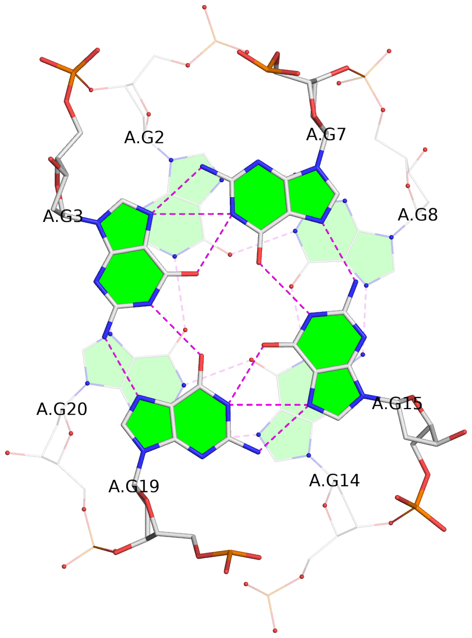
|
2 glyco-bond=-s-s sugar=---3 groove=wnwn WC-->Major O- nts=4 GGGG A.DG2,A.DG20,A.DG14,A.DG8 |
List of 1 G4-stem
In DSSR, a G4-stem is defined as a G4-helix with backbone connectivity. Bulges are also allowed along each of the four strands.
Stem#1, 3 G-tetrads, 3 loops, INTRA-molecular, UDUD, anti-parallel:chair, 2+2, 3(+Ln+Lw+Ln)
|
1 glyco-bond=s-s- sugar=-.-- groove=wnwn Major-->WC O+ nts=4 GGGG A.DG1,A.DG21,A.DG13,A.DG9 2 glyco-bond=-s-s sugar=---3 groove=wnwn WC-->Major O- nts=4 GGGG A.DG2,A.DG20,A.DG14,A.DG8 3 glyco-bond=-s-s sugar=3--- groove=wnwn WC-->Major O- nts=4 GGGG A.DG3,A.DG19,A.DG15,A.DG7 step#1 mm(<>,outward) area=12.07 rise=3.46 twist=18.4 step#2 pm(>>,forward) area=9.90 rise=3.34 twist=32.3 strand#1 U DNA glyco-bond=s-- sugar=--3 nts=3 GGG A.DG1,A.DG2,A.DG3 strand#2 D DNA glyco-bond=-ss sugar=.-- nts=3 GGG A.DG21,A.DG20,A.DG19 strand#3 U DNA glyco-bond=s-- sugar=--- nts=3 GGG A.DG13,A.DG14,A.DG15 strand#4 D DNA glyco-bond=-ss sugar=-3- nts=3 GGG A.DG9,A.DG8,A.DG7 loop#1 type=lateral strands=[#1,#4] nts=3 TTA A.DT4,A.DT5,A.DA6 loop#2 type=lateral strands=[#4,#3] nts=3 TTA A.DT10,A.DT11,A.DA12 loop#3 type=lateral strands=[#3,#2] nts=3 TTT A.DT16,A.DT17,A.DT18 |