DSSR-enabled G4 identification/annotation: PDB entry 6neb
Summary information and primary citation
- PDB-id
- 6neb
- Class
- DNA
- Method
- NMR
- Summary
- Myc promoter g-quadruplex with 1:6:1 loop length
- Reference
- Dickerhoff J, Onel B, Chen L, Chen Y, Yang D (2019): "Solution Structure of a MYC Promoter G-Quadruplex with 1:6:1 Loop Length." Acs Omega, 4, 2533-2539. doi: 10.1021/acsomega.8b03580.
- Abstract
- The important <i>MYC</i> oncogene is deregulated in many cancer cells and comprises one of the most prominent G-quadruplex (G4) forming sequences in its promoter regions, the NHE III<sub>1</sub> motif. Formation of G4s suppresses <i>MYC</i> transcription and can be modulated by drug binding, establishing these DNA structures as promising targets in cancer therapy. The NHE III<sub>1</sub> motif can fold into more than one parallel G4s, including 1:2:1 and 1:6:1 loop length conformers, with the 1:2:1 conformer shown as the major species under physiological conditions in solution. However, additional factors such as protein interactions may affect the cellular folding equilibrium. Nucleolin, a protein shown to bind <i>MYC</i> G4 and repress <i>MYC</i> transcription, is reported herein to preferably bind to the 1:6:1 loop length conformer suggesting a physiological significance of this species. The high-resolution NMR solution structure of the 1:6:1 conformer is determined, which reveals a 5'-capping structure distinctive from the 1:2:1 form, with the 6 nt central loop playing an essential role for this specific capping structure. This suggests that each parallel G-quadruplex likely adopts unique capping and loop structures determined by the specific central loop and flanking sequences. The resulting structural information at the molecular level will help to understand protein recognition of different G4s, contribution of G4 polymorphism to gene regulation, and to rationally design small molecules selectively targeting the 1:6:1 <i>MYC</i> G4.
- G4 notes
- 3 G-tetrads, 1 G4 helix, 1 G4 stem, UUUU, parallel, 4+0, 3(-P-P-P)
Cartoon-block schematics in six views (download the tarball)

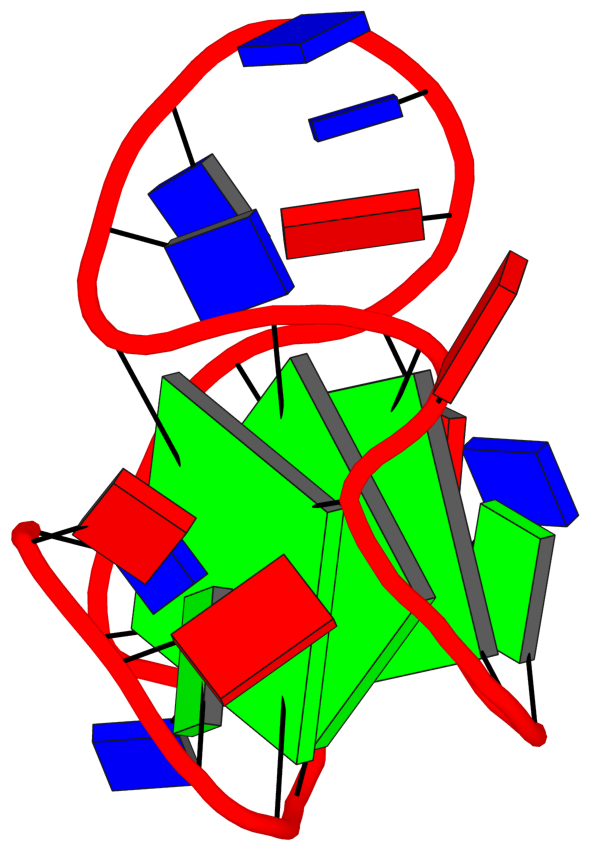
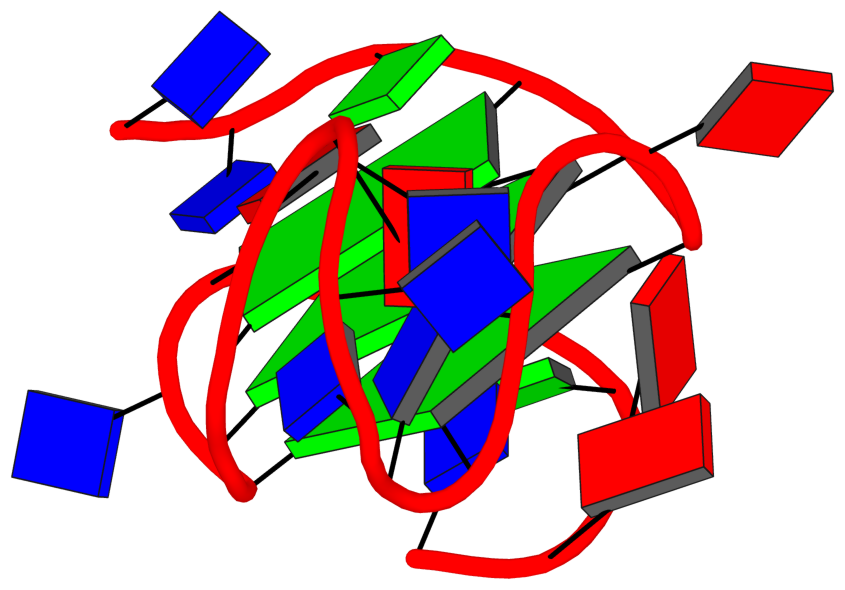
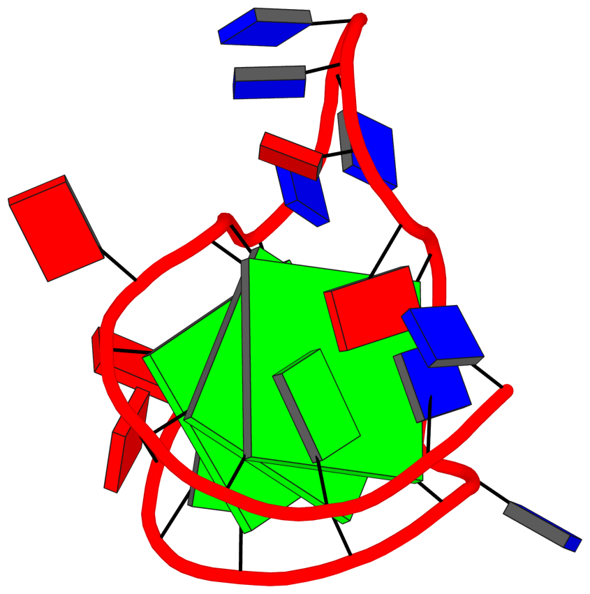
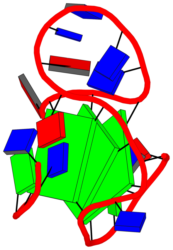
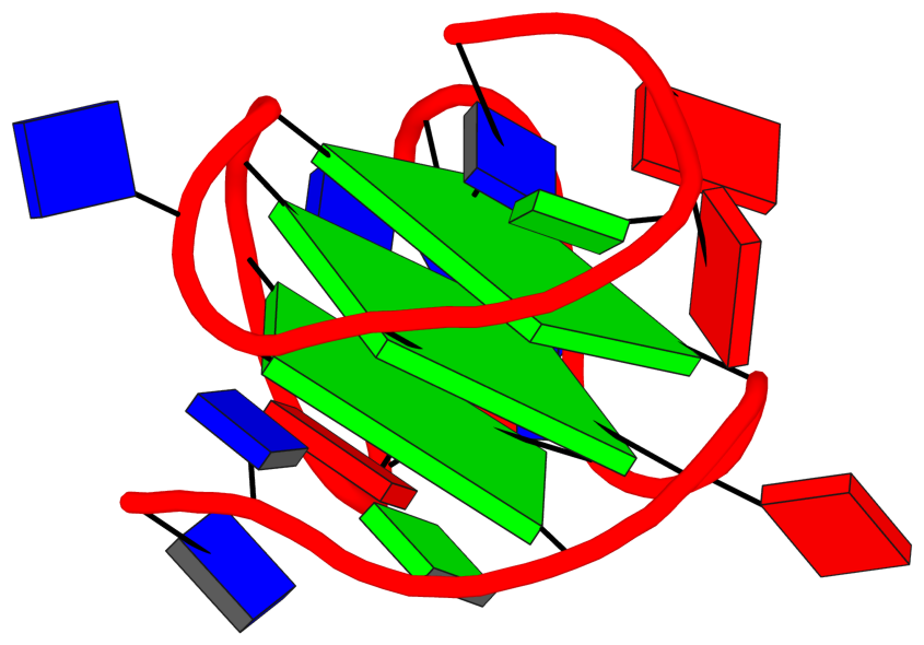
List of 3 G-tetrads
 1 glyco-bond=---- sugar=---- groove=---- planarity=0.128 type=planar O+ nts=4 GGGG A.DG3,A.DG7,A.DG16,A.DG20
 2 glyco-bond=---- sugar=---- groove=---- planarity=0.160 type=bowl-2 O+ nts=4 GGGG A.DG4,A.DG8,A.DG17,A.DG21
 3 glyco-bond=---- sugar=---- groove=---- planarity=0.343 type=bowl   O+ nts=4 GGGG A.DG5,A.DG9,A.DG18,A.DG22
List of 1 G4-helix
In DSSR, a G4-helix is defined by stacking interactions of G-tetrads, regardless of backbone connectivity, and may contain more than one G4-stem.
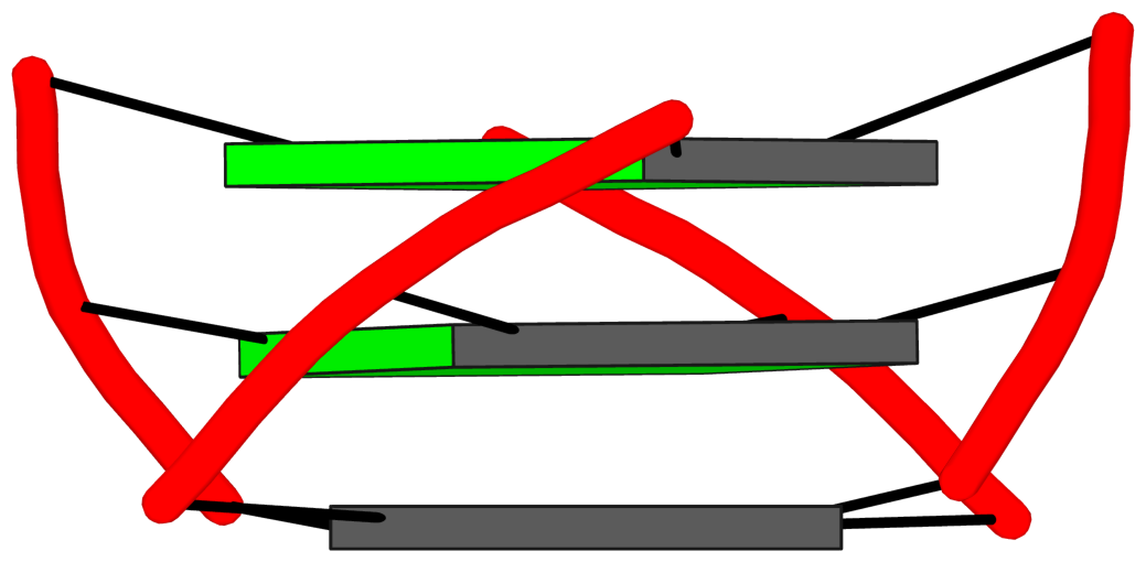
Helix#1, 3 G-tetrads, INTRA-molecular, with 1 stem
|
1 glyco-bond=---- sugar=---- groove=---- WC-->Major O+ nts=4 GGGG A.DG3,A.DG7,A.DG16,A.DG20 2 glyco-bond=---- sugar=---- groove=---- WC-->Major O+ nts=4 GGGG A.DG4,A.DG8,A.DG17,A.DG21 3 glyco-bond=---- sugar=---- groove=---- WC-->Major O+ nts=4 GGGG A.DG5,A.DG9,A.DG18,A.DG22 step#1 pm(>>,forward) area=14.54 rise=3.36 twist=24.7 step#2 pm(>>,forward) area=9.67 rise=3.48 twist=30.3 strand#1 DNA glyco-bond=--- sugar=--- nts=3 GGG A.DG3,A.DG4,A.DG5 strand#2 DNA glyco-bond=--- sugar=--- nts=3 GGG A.DG7,A.DG8,A.DG9 strand#3 DNA glyco-bond=--- sugar=--- nts=3 GGG A.DG16,A.DG17,A.DG18 strand#4 DNA glyco-bond=--- sugar=--- nts=3 GGG A.DG20,A.DG21,A.DG22 |
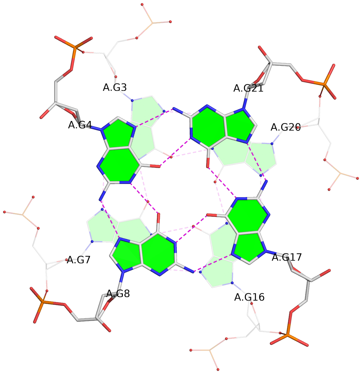
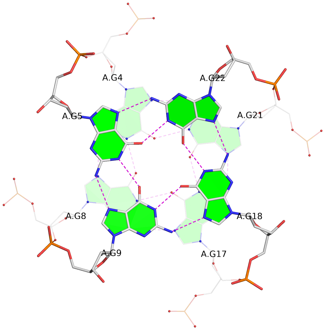
| 2 stacking diagrams | |
|
1 glyco-bond=---- sugar=---- groove=---- WC-->Major O+ nts=4 GGGG A.DG3,A.DG7,A.DG16,A.DG20 |
|
2 glyco-bond=---- sugar=---- groove=---- WC-->Major O+ nts=4 GGGG A.DG4,A.DG8,A.DG17,A.DG21 |
List of 1 G4-stem
In DSSR, a G4-stem is defined as a G4-helix with backbone connectivity. Bulges are also allowed along each of the four strands.
Stem#1, 3 G-tetrads, 3 loops, INTRA-molecular, UUUU, parallel, 4+0, 3(-P-P-P)
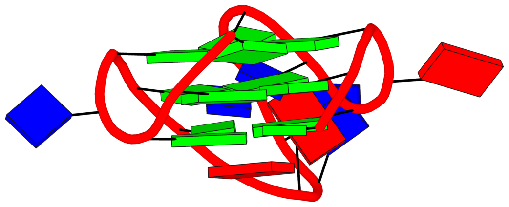
|
1 glyco-bond=---- sugar=---- groove=---- WC-->Major O+ nts=4 GGGG A.DG3,A.DG7,A.DG16,A.DG20 2 glyco-bond=---- sugar=---- groove=---- WC-->Major O+ nts=4 GGGG A.DG4,A.DG8,A.DG17,A.DG21 3 glyco-bond=---- sugar=---- groove=---- WC-->Major O+ nts=4 GGGG A.DG5,A.DG9,A.DG18,A.DG22 step#1 pm(>>,forward) area=14.54 rise=3.36 twist=24.7 step#2 pm(>>,forward) area=9.67 rise=3.48 twist=30.3 strand#1 U DNA glyco-bond=--- sugar=--- nts=3 GGG A.DG3,A.DG4,A.DG5 strand#2 U DNA glyco-bond=--- sugar=--- nts=3 GGG A.DG7,A.DG8,A.DG9 strand#3 U DNA glyco-bond=--- sugar=--- nts=3 GGG A.DG16,A.DG17,A.DG18 strand#4 U DNA glyco-bond=--- sugar=--- nts=3 GGG A.DG20,A.DG21,A.DG22 loop#1 type=propeller strands=[#1,#2] nts=1 A A.DA6 loop#2 type=propeller strands=[#2,#3] nts=6 TTTTAA A.DT10,A.DT11,A.DT12,A.DT13,A.DA14,A.DA15 loop#3 type=propeller strands=[#3,#4] nts=1 T A.DT19 |