DSSR-enabled G4 identification/annotation: PDB entry 7otb
Summary information and primary citation
- PDB-id
- 7otb
- Class
- DNA
- Method
- X-ray (1.6 Å)
- Summary
- Ruthenium polypridyl complex bound to a unimolecular chair-form g-quadruplex
- Reference
- McQuaid KT, Takahashi S, Baumgaertner L, Cardin DJ, Paterson NG, Hall JP, Sugimoto N, Cardin CJ (2022): "Ruthenium Polypyridyl Complex Bound to a Unimolecular Chair-Form G-Quadruplex." J.Am.Chem.Soc., 144, 5956-5964. doi: 10.1021/jacs.2c00178.
- Abstract
- The DNA G-quadruplex is known for forming a range of topologies and for the observed lability of the assembly, consistent with its transient formation in live cells. The stabilization of a particular topology by a small molecule is of great importance for therapeutic applications. Here, we show that the ruthenium complex Λ-[Ru(phen)<sub>2</sub>(qdppz)]<sub>2+</sub> displays enantiospecific G-quadruplex binding. It crystallized in 1:1 stoichiometry with a modified human telomeric G-quadruplex sequence, GGGTTAGGGTTAGGGTTTGGG (<i>htel21</i>T<sub>18</sub>), in an antiparallel chair topology, the first structurally characterized example of ligand binding to this topology. The lambda complex is bound in an intercalation cavity created by a terminal G-quartet and the central narrow lateral loop formed by T<sub>10</sub>-T<sub>11</sub>-A<sub>12</sub>. The two remaining wide lateral loops are linked through a third K<sub>+</sub> ion at the other end of the G-quartet stack, which also coordinates three thymine residues. In a comparative ligand-binding study, we showed, using a Klenow fragment assay, that this complex is the strongest observed inhibitor of replication, both using the native human telomeric sequence and the modified sequence used in this work.
- G4 notes
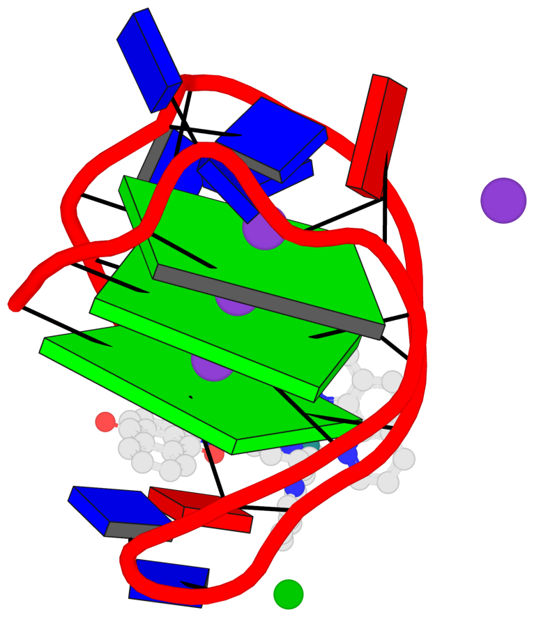
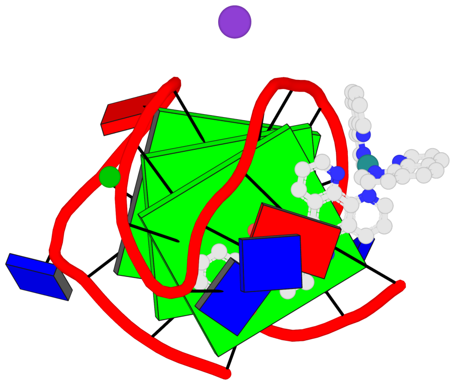
- 3 G-tetrads, 1 G4 helix, 1 G4 stem, UDUD, anti-parallel:chair, 2+2, 3(-Lw-Ln-Lw)
Cartoon-block schematics in six views (download the tarball)
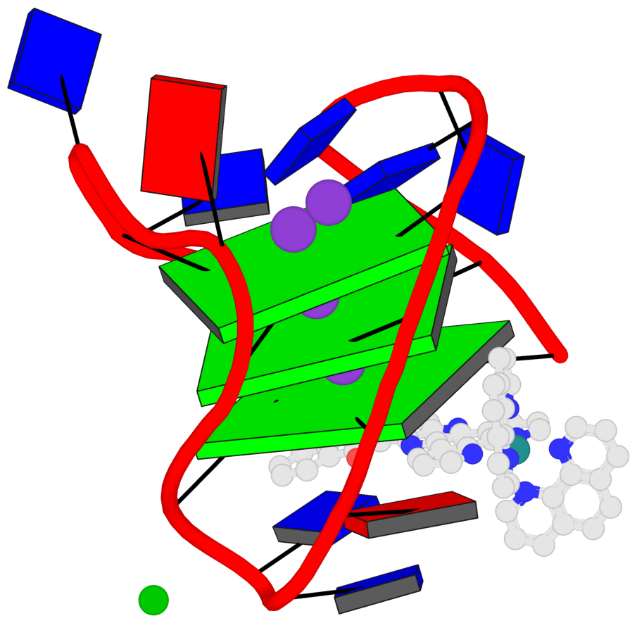
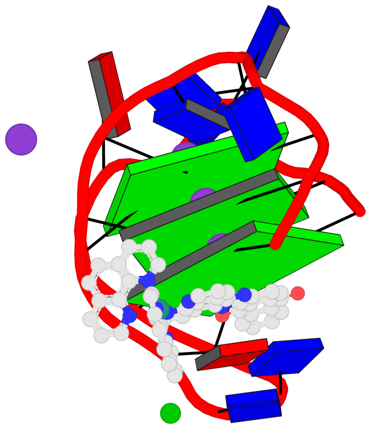
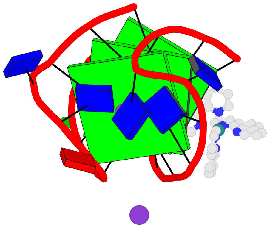
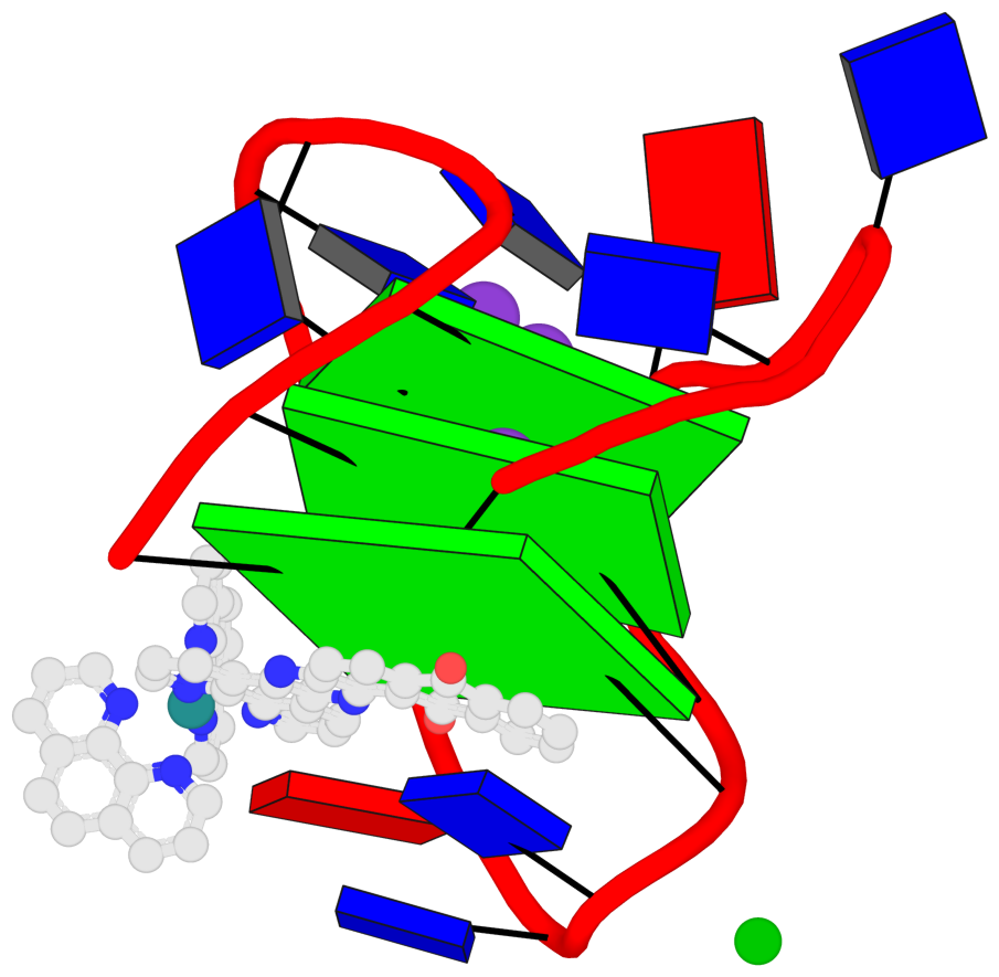
List of 3 G-tetrads
 1 glyco-bond=s-s- sugar=---- groove=wnwn planarity=0.543 type=bowl-2 O- nts=4 GGGG A.DG1,A.DG9,A.DG13,A.DG21
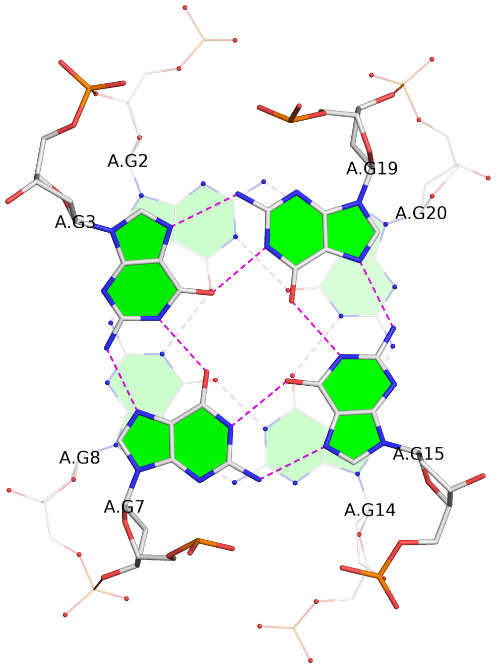
 2 glyco-bond=s-s- sugar=--.- groove=wnwn planarity=0.515 type=other  O- nts=4 GGGG A.DG2,A.DG8,A.DG14,A.DG20
 3 glyco-bond=-s-s sugar=---- groove=wnwn planarity=0.568 type=saddle O+ nts=4 GGGG A.DG3,A.DG7,A.DG15,A.DG19
List of 1 G4-helix
In DSSR, a G4-helix is defined by stacking interactions of G-tetrads, regardless of backbone connectivity, and may contain more than one G4-stem.
Helix#1, 3 G-tetrads, INTRA-molecular, with 1 stem
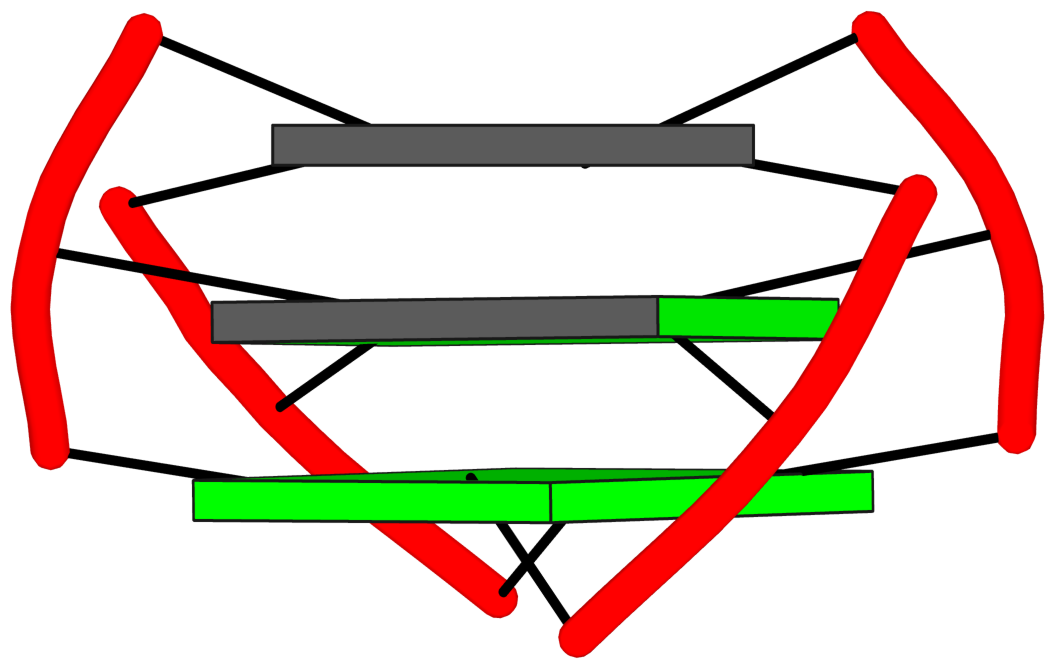
|
1 glyco-bond=s-s- sugar=---- groove=wnwn Major-->WC O- nts=4 GGGG A.DG1,A.DG9,A.DG13,A.DG21 2 glyco-bond=s-s- sugar=--.- groove=wnwn Major-->WC O- nts=4 GGGG A.DG2,A.DG8,A.DG14,A.DG20 3 glyco-bond=-s-s sugar=---- groove=wnwn WC-->Major O+ nts=4 GGGG A.DG3,A.DG7,A.DG15,A.DG19 step#1 mp(<<,backward) area=17.44 rise=3.51 twist=22.0 step#2 mm(<>,outward) area=12.49 rise=3.50 twist=20.0 strand#1 DNA glyco-bond=ss- sugar=--- nts=3 GGG A.DG1,A.DG2,A.DG3 strand#2 DNA glyco-bond=--s sugar=--- nts=3 GGG A.DG9,A.DG8,A.DG7 strand#3 DNA glyco-bond=ss- sugar=-.- nts=3 GGG A.DG13,A.DG14,A.DG15 strand#4 DNA glyco-bond=--s sugar=--- nts=3 GGG A.DG21,A.DG20,A.DG19 |
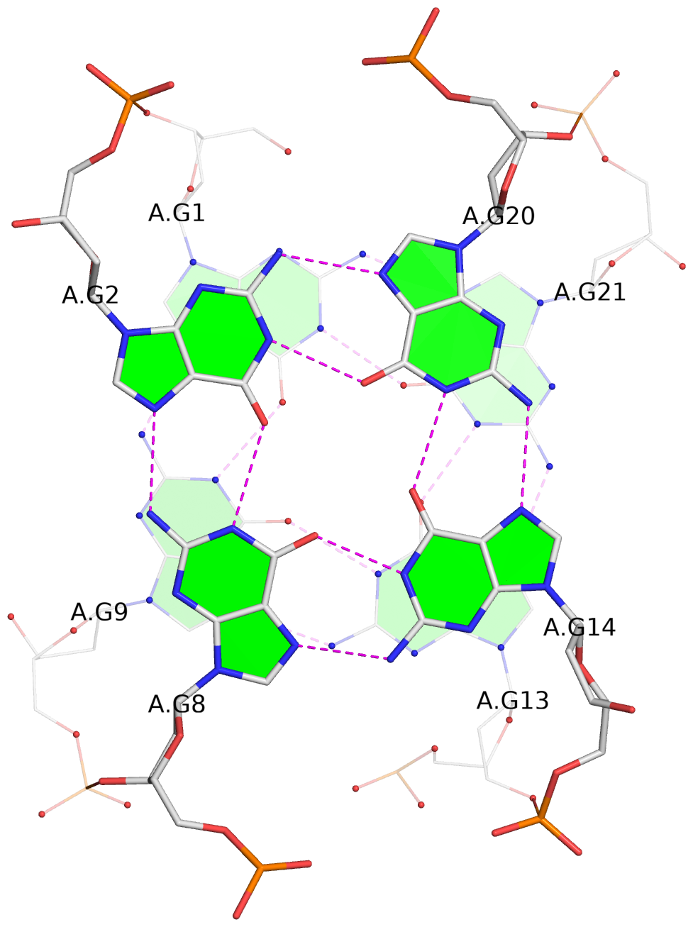
| 2 stacking diagrams | |
|
1 glyco-bond=s-s- sugar=---- groove=wnwn Major-->WC O- nts=4 GGGG A.DG1,A.DG9,A.DG13,A.DG21 |
|
2 glyco-bond=s-s- sugar=--.- groove=wnwn Major-->WC O- nts=4 GGGG A.DG2,A.DG8,A.DG14,A.DG20 |
List of 1 G4-stem
In DSSR, a G4-stem is defined as a G4-helix with backbone connectivity. Bulges are also allowed along each of the four strands.
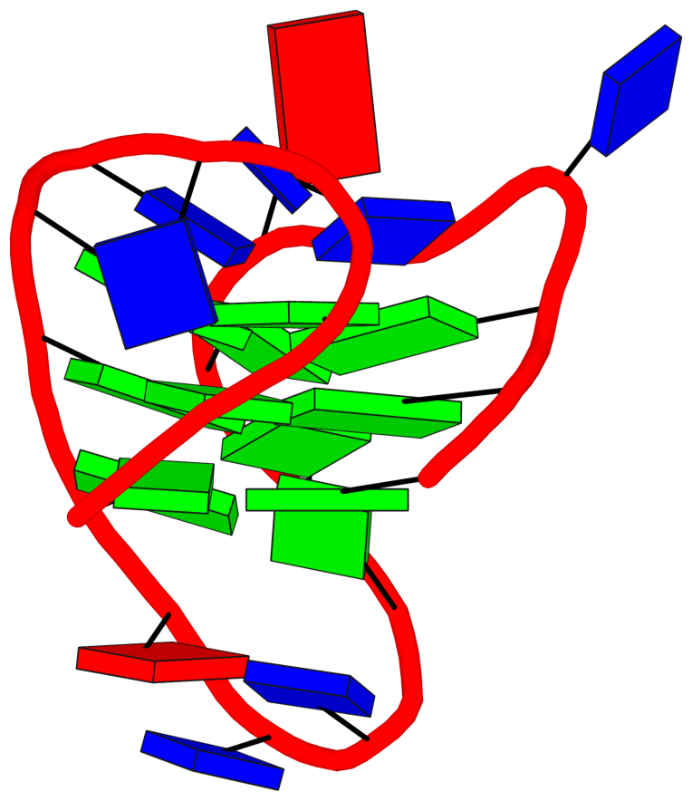
Stem#1, 3 G-tetrads, 3 loops, INTRA-molecular, UDUD, anti-parallel:chair, 2+2, 3(-Lw-Ln-Lw)
|
1 glyco-bond=s-s- sugar=---- groove=wnwn Major-->WC O- nts=4 GGGG A.DG1,A.DG9,A.DG13,A.DG21 2 glyco-bond=s-s- sugar=--.- groove=wnwn Major-->WC O- nts=4 GGGG A.DG2,A.DG8,A.DG14,A.DG20 3 glyco-bond=-s-s sugar=---- groove=wnwn WC-->Major O+ nts=4 GGGG A.DG3,A.DG7,A.DG15,A.DG19 step#1 mp(<<,backward) area=17.44 rise=3.51 twist=22.0 step#2 mm(<>,outward) area=12.49 rise=3.50 twist=20.0 strand#1 U DNA glyco-bond=ss- sugar=--- nts=3 GGG A.DG1,A.DG2,A.DG3 strand#2 D DNA glyco-bond=--s sugar=--- nts=3 GGG A.DG9,A.DG8,A.DG7 strand#3 U DNA glyco-bond=ss- sugar=-.- nts=3 GGG A.DG13,A.DG14,A.DG15 strand#4 D DNA glyco-bond=--s sugar=--- nts=3 GGG A.DG21,A.DG20,A.DG19 loop#1 type=lateral strands=[#1,#2] nts=3 TTA A.DT4,A.DT5,A.DA6 loop#2 type=lateral strands=[#2,#3] nts=3 TTA A.DT10,A.DT11,A.DA12 loop#3 type=lateral strands=[#3,#4] nts=3 TTT A.DT16,A.DT17,A.DT18 |