DSSR-enabled G4 identification/annotation: PDB entry 9gxh
Summary information and primary citation
- PDB-id
- 9gxh
- Class
- immune system
- Method
- X-ray (1.9 Å)
- Summary
- Nanobody bound to tba g-quadruplex
- Reference
- Pevec M, Medved T, Kovacic M, Zerjav N, Imperl J, Plavec J, Lah J, Loris R, Hadzi S (2025): "Structural basis of G-quadruplex recognition by a camelid antibody fragment." Nucleic Acids Res., 53. doi: 10.1093/nar/gkaf453.
- Abstract
- Apart from the iconic Watson-Crick duplex, DNA can fold into different noncanonical structures, of which the most studied are G-quadruplexes (G4s). Despite mounting structural and biophysical evidence, their existence in cells was controversial until their detection using G4-specific antibodies. However, it remains unknown how antibodies recognize G4s at the molecular level and why G4-specific antibodies have low selectivity and are unable to distinguish different G4 sequences. Here, we present the crystal structure of a nanobody bound to the archetypical G4 structure, the thrombin-binding aptamer (TBA). The nanobody exhibits strong selectivity against different G4 sequences and utilizes an unusual scaffold-based paratope, with very limited involvement of complementarity-determining region. The nanobody effectively mimics the binding interface of thrombin, a natural binding partner of TBA, by using isosteric interactions at key positions. The presented structure sheds light on the molecular basis of how antibodies, essential G4-detection tools, recognize noncanonical G4 structures.
- G4 notes
- 2 G-tetrads, 1 G4 helix, 1 G4 stem, UDUD, anti-parallel:chair, 2+2, 2(+Ln+Lw+Ln)
Cartoon-block schematics in six views (download the tarball)
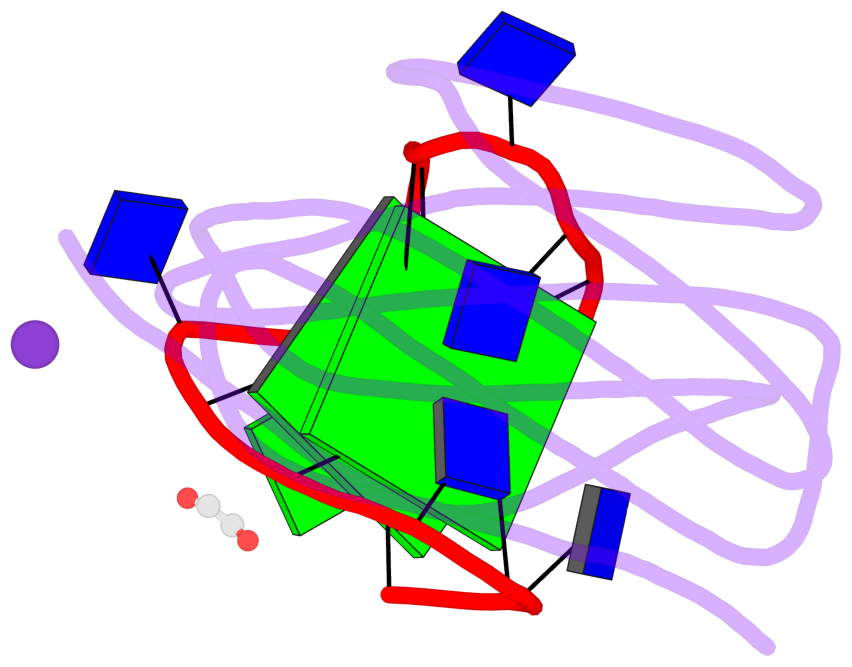
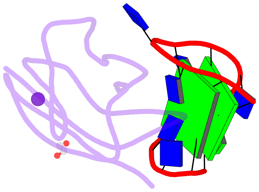
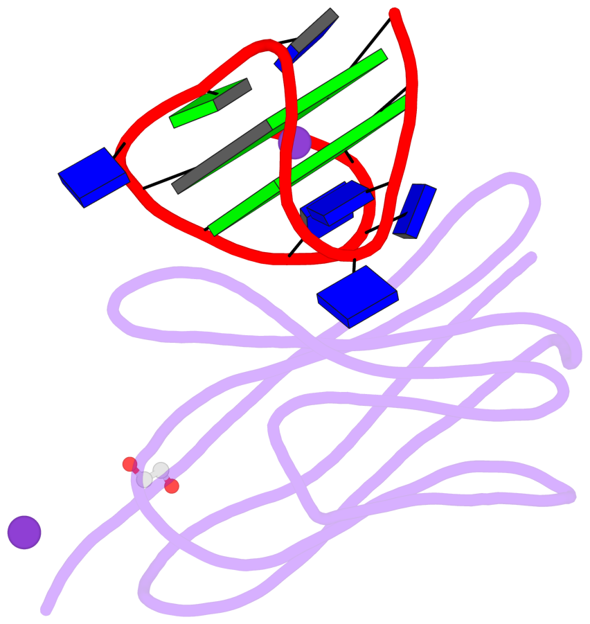
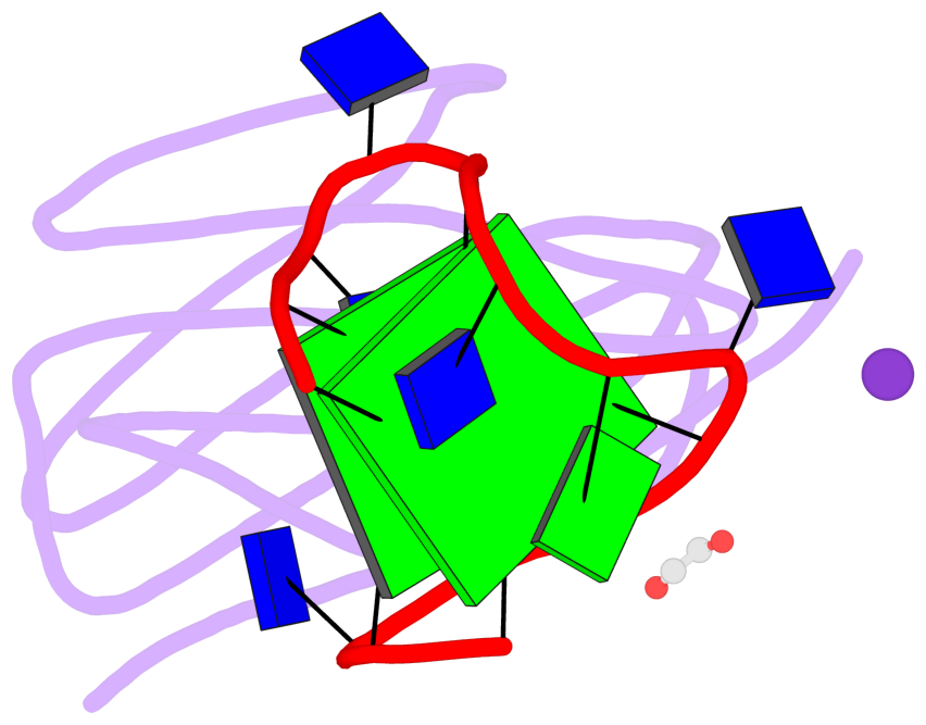
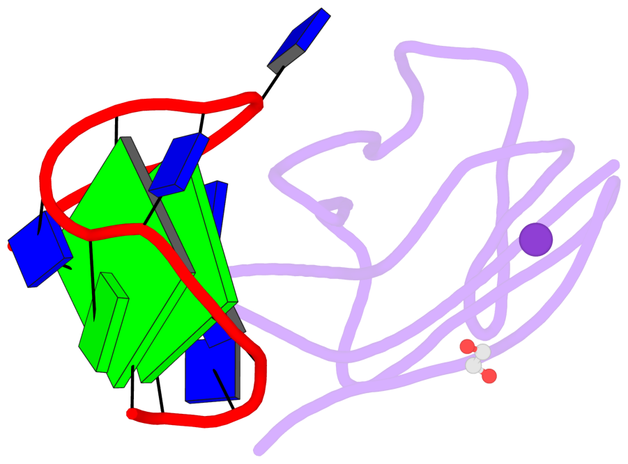
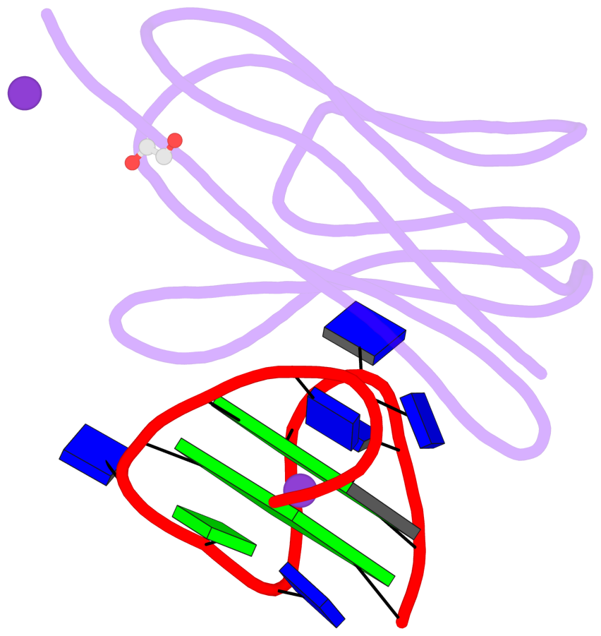
List of 2 G-tetrads
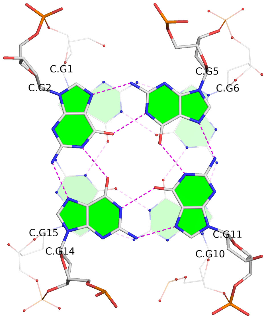
 1 glyco-bond=s-s- sugar=---- groove=wnwn planarity=0.331 type=bowl   O+ nts=4 GGGG 2:C.DG1,2:C.DG15,2:C.DG10,2:C.DG6
 2 glyco-bond=-s-s sugar=.--- groove=wnwn planarity=0.249 type=bowl-2 O- nts=4 GGGG 2:C.DG2,2:C.DG14,2:C.DG11,2:C.DG5
List of 1 G4-helix
In DSSR, a G4-helix is defined by stacking interactions of G-tetrads, regardless of backbone connectivity, and may contain more than one G4-stem.
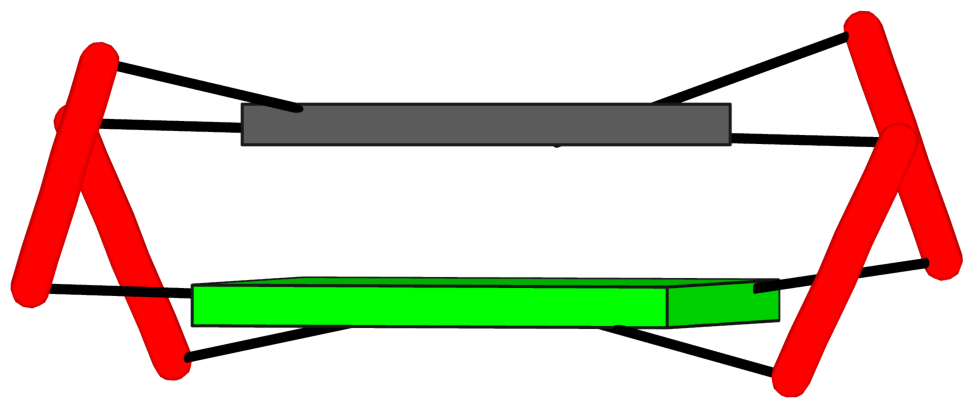
Helix#1, 2 G-tetrads, INTRA-molecular, with 1 stem
|
1 glyco-bond=s-s- sugar=---- groove=wnwn Major-->WC O+ nts=4 GGGG 2:C.DG1,2:C.DG15,2:C.DG10,2:C.DG6 2 glyco-bond=-s-s sugar=.--- groove=wnwn WC-->Major O- nts=4 GGGG 2:C.DG2,2:C.DG14,2:C.DG11,2:C.DG5 step#1 mm(<>,outward) area=17.98 rise=3.51 twist=13.2 strand#1 DNA glyco-bond=s- sugar=-. nts=2 GG 2:C.DG1,2:C.DG2 strand#2 DNA glyco-bond=-s sugar=-- nts=2 GG 2:C.DG15,2:C.DG14 strand#3 DNA glyco-bond=s- sugar=-- nts=2 GG 2:C.DG10,2:C.DG11 strand#4 DNA glyco-bond=-s sugar=-- nts=2 GG 2:C.DG6,2:C.DG5 |
| 1 stacking diagram | |
|
1 glyco-bond=s-s- sugar=---- groove=wnwn Major-->WC O+ nts=4 GGGG 2:C.DG1,2:C.DG15,2:C.DG10,2:C.DG6 |
List of 1 G4-stem
In DSSR, a G4-stem is defined as a G4-helix with backbone connectivity. Bulges are also allowed along each of the four strands.
Stem#1, 2 G-tetrads, 3 loops, INTRA-molecular, UDUD, anti-parallel:chair, 2+2, 2(+Ln+Lw+Ln)
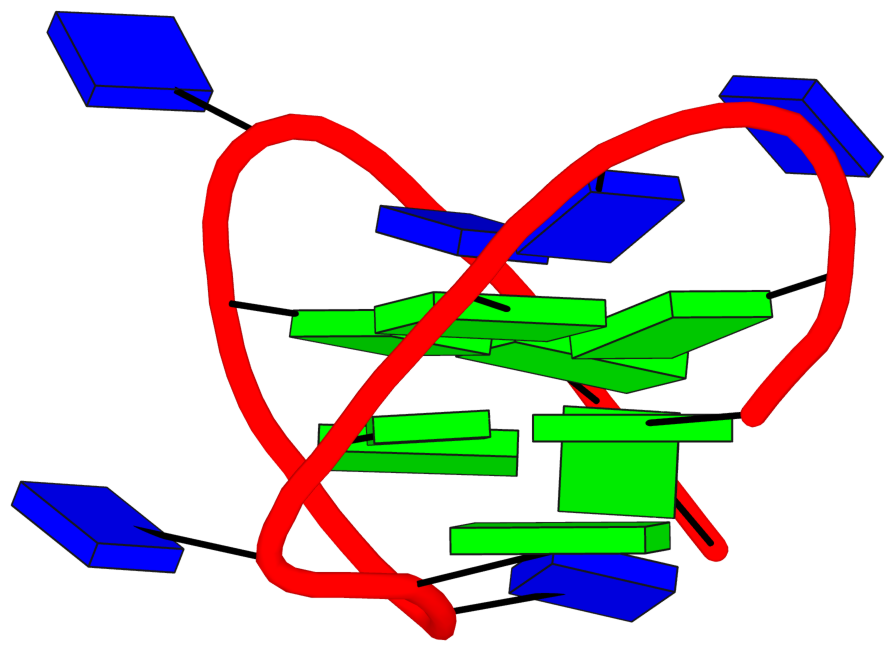
|
1 glyco-bond=s-s- sugar=---- groove=wnwn Major-->WC O+ nts=4 GGGG 2:C.DG1,2:C.DG15,2:C.DG10,2:C.DG6 2 glyco-bond=-s-s sugar=.--- groove=wnwn WC-->Major O- nts=4 GGGG 2:C.DG2,2:C.DG14,2:C.DG11,2:C.DG5 step#1 mm(<>,outward) area=17.98 rise=3.51 twist=13.2 strand#1 U DNA glyco-bond=s- sugar=-. nts=2 GG 2:C.DG1,2:C.DG2 strand#2 D DNA glyco-bond=-s sugar=-- nts=2 GG 2:C.DG15,2:C.DG14 strand#3 U DNA glyco-bond=s- sugar=-- nts=2 GG 2:C.DG10,2:C.DG11 strand#4 D DNA glyco-bond=-s sugar=-- nts=2 GG 2:C.DG6,2:C.DG5 loop#1 type=lateral strands=[#1,#4] nts=2 TT 2:C.DT3,2:C.DT4 loop#2 type=lateral strands=[#4,#3] nts=3 TGT 2:C.DT7,2:C.DG8,2:C.DT9 loop#3 type=lateral strands=[#3,#2] nts=2 TT 2:C.DT12,2:C.DT13 |